Figure 3.
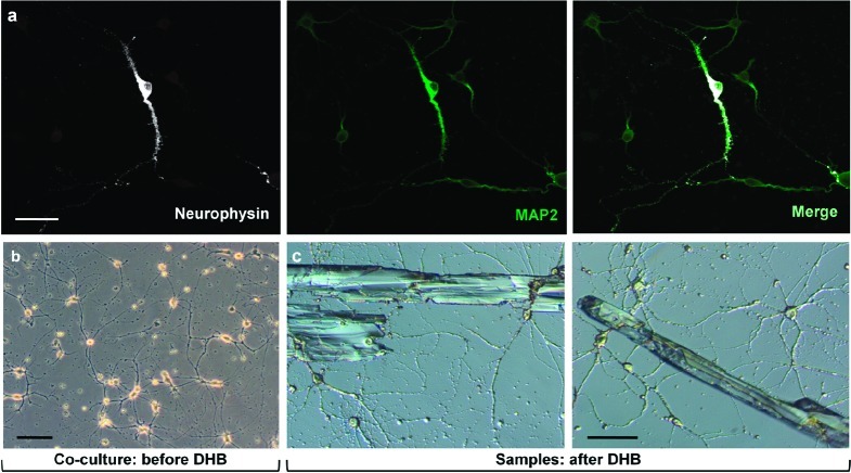
Magnocellular neuron development is improved by hippocampal neurons in coculture. (a) Magnocellular neurons were plated onto a 5-day-old culture of hippocampal neurons and maintained for 7 days. Magnocellular neurons show abundant NP (white), while hippocampal neurons lack NP. MAP2 content (green) of the neurosecretory magnocellular neuron is abundant, compared with hippocampal neurons. (b) Low magnification, phase-contrast image of primary magnocellular neuron−hippocampal neuron coculture demonstrates neuronal connectivity. (c) After cultures were rapidly rinsed with DHB, crystals form to embed neurons and locally extract peptides (differential interference contrast microscopy). For this type of MS analysis, neuronal cells must be embedded in matrix crystals. Samples were dried for 1−2 days in a desiccator prior to performance of MALDI analysis. All scale bars = 50 μm.

