
Figure 5.
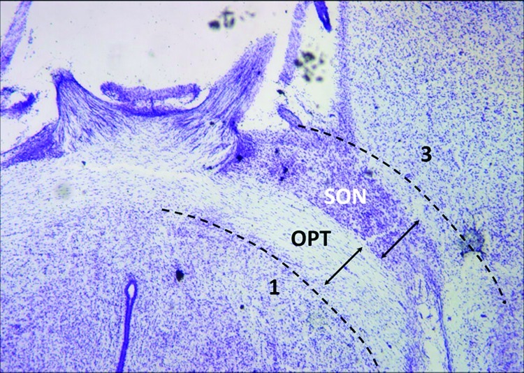
Horizontal hypothalamic brain slice of postnatal rat. Using the optic tracts (OPT) as landmarks, four dissection steps were used to remove the tissue containing the supraoptic nucleus (SON). (1) Posterior cuts (shown): bilateral incisions were made along the immediate posterior surface of the optic tract to a point where the tracts enter into the brain and are no longer visible from the ventral surface. (2) Lateral cuts (not shown): one cut per side of the brain to sever the optic tract where it penetrates into the brain near the rostral cerebral artery. (3) Anterior cuts (shown): these incisions were performed away from the surface of the optic tract, by about the same width of the OPT, to ensure that the supraoptic nucleus would be retained and not damaged in the cutting. (4) Dorsal cut (not shown): this last cut is made as close to the dorsal border of the optic tract as possible. Upon completion of these four steps, the excised tissue containing the supraoptic nucleus and a portion of the optic tract is removed. The images are paraformaldehyde-fixed sections from a single postnatal rat brain, sectioned horizontally by a cryostat and histochemically stained with cresyl violet to identify the Nissl substance. The supraoptic nucleus appears as a darkly stained cluster of cells adjacent to the optic tracts.