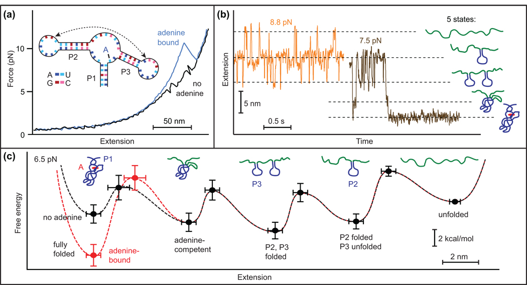
Figure 3.
SMFS of folding in an adenine riboswitch aptamer. (a) Binding of the adenine ligand alters the shape of the FECs. (b) Constant-force trajectories obtained at two different forces indicate five distinct states in the folding, corresponding to the formation of specific structural elements of the aptamer, as indicated. (c) Energy landscape for the complete folding reaction, both with (red) and without (black) ligand. Adapted from [26••].

