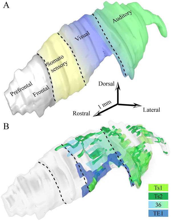
Figure 6.
Sensory sectors of the rhesus monkey TRN. A, Three-dimensional reconstruction of TRN (light gray) showing the approximate sectors receiving projections from auditory (green), visual (blue) and somatosensory (yellow) cortices in non-human primates based on available evidence. The colors between the different sectors change gradually to illustrate the blurred topography of the sectors and their overlaps. B, Three-dimensional reconstruction of TRN (light gray), which was rendered transparent to show the position of axonal terminations from temporal sensory association cortices, including auditory areas Ts1 (light green) and Ts2 (dark green), visual area TE1 (dark blue), and polymodal area 36 (light green-blue gradient).

