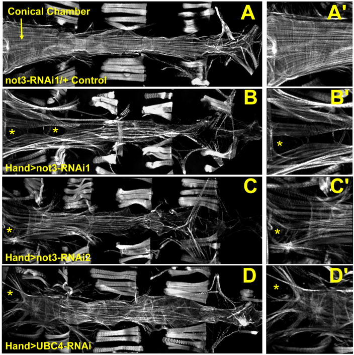
Figure 4. not3 and UBC4 cardiac specific RNAi-knockdown substantially perturb myofibrillar organization and content.
(A) Alexa584-phalloidin staining of control Drosophila cardiac tubes reveals typical spiraling myofibrillar arrangements within the cardiomyocytes. The fibers, especially those in the conical chamber, located anteriorly, are densely packed with f-actin. (B–D) Relative to control hearts, not3 or UBC4 RNAi knockdown severely disrupts myofibrillar organization and leads to an apparent loss of myofilaments as noted by large gaps in f-actin staining (*) as well as by a lack of myosin heavy chain transcripts (Fig. S4F). (A′-D′) Enlarged images of the conical chambers from A–D, respectively, which illustrate the high degree of myofibrillar disarray and large gaps in f-actin staining within the cardiomyocytes of not3 and UBC4 RNAi knockdown hearts. Original images taken at 10X magnification with a Zeiss Imager Z1 fluorescent microscope.

