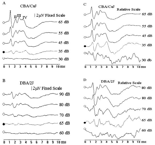
Fig. 1.
Determination of ABR thresholds. Normal ABR patterns for a 32 week old CBA/CaJ mouse (A) induced by click stimuli of 65, 55, 45, 35 and 30 dB SPL. The ABR threshold for this mouse was estimated to be 35 dB SPL. (There are usually 4 or 5 response peaks, labeled I, II, II, and IV respectively.) ABR patterns for a 5 week old DBA/2J mouse (B) induced by click stimuli of 90, 80, 70, 65 and 60dB SPL. The ABR threshold for this mouse was estimated to be 65 dB SPL. Response amplitudes for all four stimuli are shown on the same fixed scale in A and B to better compare the amplitude differences between normal and impaired mice. For routine threshold determinations, ABR were displayed in a relative scale adjusted to maximize the wave pattern for each stimulus as shown in C and D, because these wave patterns were more easily recognized thans when displayed in a fixed scale.

