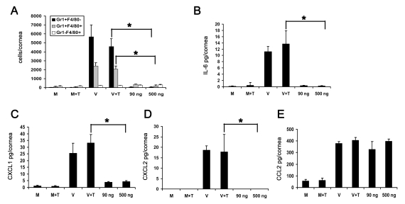
Figure 5. Adenoviral genomic DNA induces differential expression of cytokines but does not cause infiltration of leukocytes into the cornea.
(A) Flow cytometric analysis of Gr1 and F4/80 positive cells in mouse corneas at 4 days after injection with virus free buffer (M), virus free buffer and transfection reagent (M+T), intact HAdV-37 (V), intact HAdV-37 and transfection reagent (V+T), and 90 ng or 500 ng of HAdV-37 genomic DNA with transfection reagent (n = 6 corneas/group). Data shown represents the mean of three independent experiments, and error bars represent SD. (B–E) Protein levels of cytokines IL-6 (B), CXCL1 (C), CXCL2 (D) and CCL2 (E) in mouse corneas at 16 hpi. Corneas were injected with virus free buffer (M), virus free buffer and transfection reagent (M+T), intact HAdV-37 (V), intact HAdV-37 and transfection reagent (V+T), and 90 ng or 500 ng of HAdV-37 genomic DNA with transfection reagent (n = 9 corneas/group). Data shown represents the mean of three separate experiments, and error bars denote SD. * p<.05, ANOVA.

