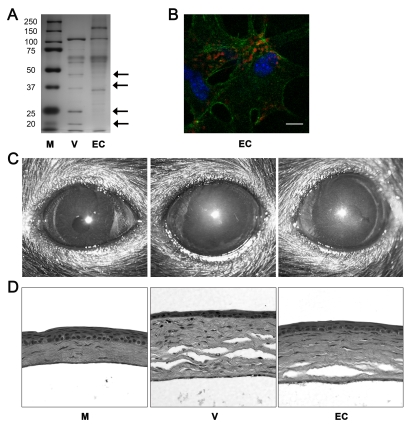
Figure 6. Empty adenoviral capsid is sufficient to induce keratitis.
(A) Silver stained polyacrylamide gel of proteins from intact HAdV-37 (V) or empty capsid (EC). First lane (M) shows protein standards. Arrows on the right point to capsid proteins missing from the empty capsid; capsid proteins V and VII are marked by the second and fourth arrows from the top, respectively. (B) Mouse cornea injected with Cy3 dye-labeled empty capsid (EC). Intracellular virus position was visualized with confocal microscopy at 90 min pi (n = 3 corneas). Red: Cy3-labeled empty capsid. Green: intracellular actin (phalloidin stain). Blue: nuclei (TO-PRO3 stain). Scale bar 20 µM. (C) Clinical appearance and (D) histopathology of mouse corneas at 4 dpi. Corneas were injected with virus free buffer (M), intact virus (V), or empty capsid (EC) (n = 5 mice/group).

