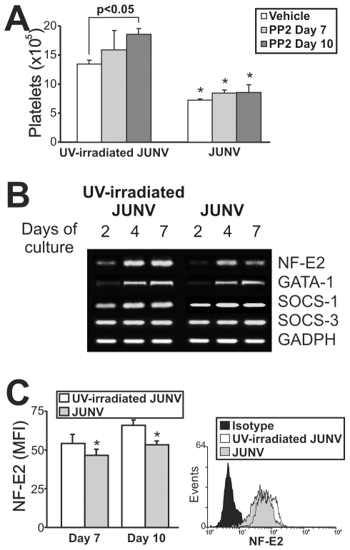
Figure 7. Intracellular mechanisms involved in the JUNV-induced inhibition of platelet production.
CD34+ cells were UV-irradiated JUNV- or JUNV-infected, washed and stimulated with TPO. (A) The Src inhibitor PP2 (10 µM) was added at the indicated days, and Plt counts were determined at culture day 15. The values represent the mean ± SEM of four independent experiments, * indicates p<0.05 vs. UV-irradiated JUNV-treated cells. (B) Semi-quantitative RT-PCR analysis of relevant molecules involved in megakaryo/thrombopoiesis were performed at the indicated days of culture. The figure shows a representative experiment of three similar replicates. (C) NF-E2 expression was assessed in the megakaryocytic population by immunostaining the cells first with a PE-conjugated anti-CD41 mAb or an isotype-matched control. Then the cells were incubated with anti-NF-E2 polyclonal followed by FITC-conjugated swine anti-rabbit Igs. Cells were analyzed by flow cytometry. Non-specific fluorescence was assessed using rabbit serum instead of primary Ab. The values represent the mean ± SEM of three independent experiments,* indicates p<0.05 vs. UV-irradiated JUNV-infected cells. The histogram shows a representative flow cytometric analysis at day ten.

