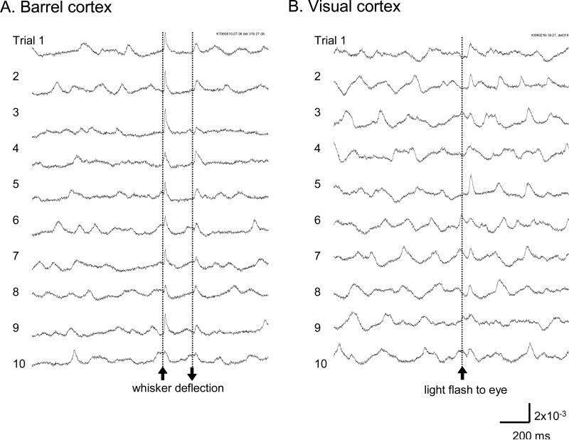
Figure 7. Sensory evoked response.
A. Signal from a single detector viewing a cortical area of 160 μm in diameter near the principal barrel in the barrel cortex. Ten trials are displayed from 105 trials with identical whisker stimuli. B. Signal from a single detector viewing a cortical area of 160 μm in diameter in the V1 area. Ten trials are displayed from ~30 trials recorded with identical light stimuli, to the contralateral eye.

