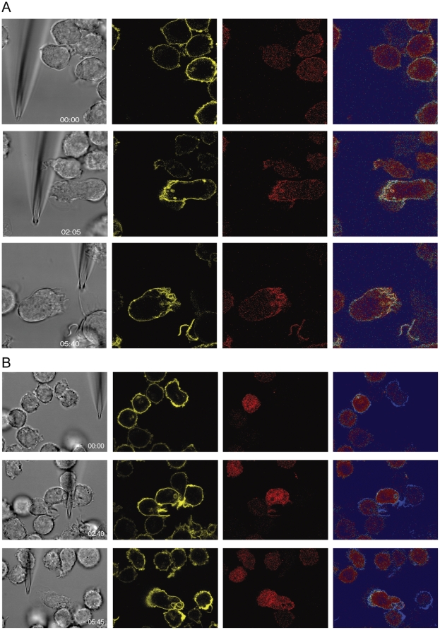
Figure 8. Role of PIP3 formation during reversal of polarization.
Selected frames from Movies S6 just after insertion of the micropipette dispensing 300 nM UK 14'304 (0 sec, upper images) and after 5:40 min (lower images). The emission of THP-1 cells transduced with PH-PKB-mCherry (red fluorescence, 594 nm excitation/610–680 nm emission) and expressing α2AAR-YFP/CFP (yellow fluorescence YFP, 514 nm excitation/525–580 nm emission) was recorded contemporaneously. The false color shows the ratio of red/yellow (right panels). Gray, corresponding phase image. (A) control cells, (B) cell pretreated for 10 min with 100 nM Wortmannin. (514 nm excitation/525–590 nm emission), (Phase) phase images taken with DIC settings, and (Ratio) False color shows the ratio of red/yellow.

