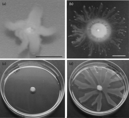
Fig. 4.
Swarming motility on LB plates. Two clinical isolates displayed colony spread consistent with swarming when assayed on 0.3 % agar LB plates. The first isolate (a) displayed no swimming, while the second (b) displayed both a swimming zone and a swarming morphology on the agar surface. Bars, 1 cm. (c) PAO1 does not swarm on 0.5 % agar LB (48 h post-inoculation), while the clinical isolate pictured in (b) shows extensive swarming on 0.5 % agar LB at this time (d).

