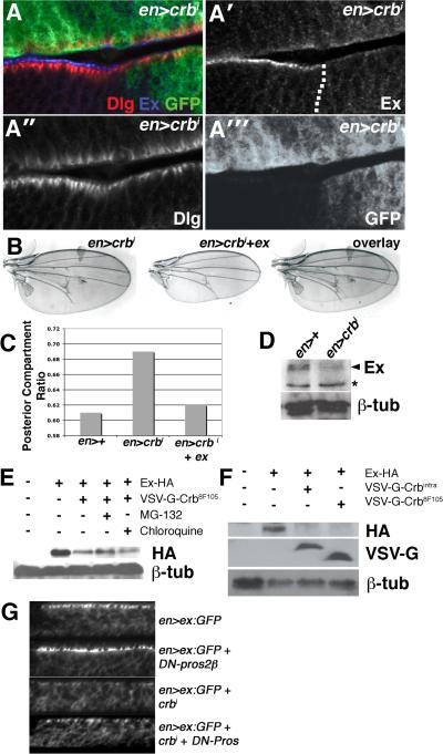
Figure 3. crbi downregulates Ex levels.
(A-A′″) Lateral section of en>crbi,GFP wing disc co-stained for Dlg (red) and Ex (blue). Dotted line denotes A:P boundary. (B) Images and overlays of en>crbi and en>crbi,ex wings. (C) PCR in the indicated genotypes. (D) Immunoblot of Ex in en>+ and en>crbi wing discs. Arrowhead denotes Ex based on comigration with overexpressed Ex (not shown) (* = non-specific band). Lower panel is α-β-tub loading control. (E) Immunoblot of HA-Ex in Crb8F105-expressing cells treated with the MG132 (lane 4) or chloroquine (lane 5). (F) Corresponding α-HA, α-VSV-G, and α-β-tub immunoblots of S2 cells expressing HA-Ex from the pAct-HA-Ex plasmid (lanes 2-4), and VSV-G-tagged forms of either crbi (lane 3) or crb8F105 (lane 4) from the pMT plasmid. (G) Lateral images of Ex:GFP in the posterior region of the wing pouch in the indicated genotypes.

