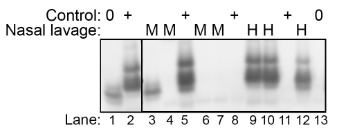
Figure 6. Western blot detection of HY TME infection in nasal lavages using the QuIC assay.
Nasal lavages (2 µl) from mock- (M)(lanes 3, 4, 6, and 7 correspond to the four mock-infected samples from Table 3) and HY TME (H) -infected hamsters (lanes 9, 10, and 12 correspond to HY TME i.ob. #1 to #3 in Table 3) were subject to two serial rounds of the QuIC assay in order to detect low levels of PrPSc. PK-resistant PrP polypeptides between 11 and 17 kDa were observed in the HY TME nasal lavage samples indicating amplification of PrPSc. In mock-infected nasal lavage samples either no polypeptides or occassionally lower molecular weight polypeptides (≤12 kDa) were observed, but the latter were not observed in prion-positive control samples. Controls include mock-infected hamster brain homogenates at a 10−2 and 10−6 dilution (lanes 1 and 13, respectively), HY TME hamster brain at a 10−6, 10−8, and 10−10 dilution (lanes 5, 8, and 11, respectively), and 100 femtogram of purified PrPSc from 263K scrapie infected hamster brain (lane 2).

