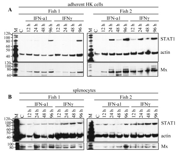
Figure 4.
SDS-PAGE followed by Western blot showing expression of STAT1 protein in (A) adherent head kidney leukocytes and (B) splenocytes from two individuals upon stimulation with IFN. The samples were harvested 12, 24, 48, and 96 h after stimulation with IFN-a1 (10 U/mL) and IFNγ (200 ng/mL). The unstimulated control, C, was harvested at the 48 h time-point. STAT1 protein was detected simultaneously with actin which was used as a loading control. The membranes were stripped and reprobed with an anti-Mx-antibody as a control for the IFN-activity. M = MagicMark molecular weight marker.

