Fig. 1.

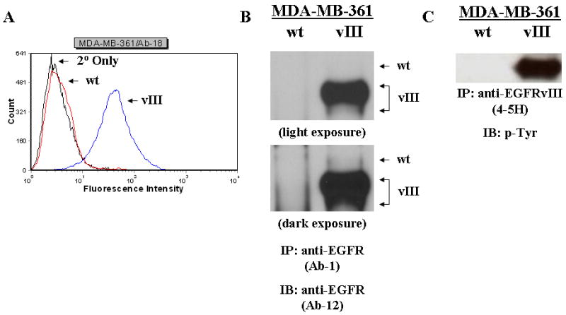
EGFRvIII expression in MDA-MB-361/vIII cells. a, the levels of EGFRvIII in the MDA-MB-361/vIII transfectants were quantitatively measured by flow cytometry using an anti-EGFRvIII antibody (Ab-18). The leftmost curve (black line) represents nonspecific staining (primary antibody omitted). The other curves represent the expression of EGFRvIII receptor in MDA-MB-361/wt (red line) and MDA-MB-361/vIII (blue line) cells. b and c, Immunoprecipitation and immunoblot analysis of EGFRvIII. Whole cell lysates (500-1000 μg) from MDA-MB-361/wt and MDA-361/vIII cells were immunoprecipitated with an anti-EGFR (Ab-1) or anti-EGFRvIII (4-5H) antibody, electrophoresed on SDS-PAGE, transferred onto nitrocellulose membranes, and immunoblotted with either an anti-EGFR (Ab-12) or anti-phosphotyrosine antibody. Protein bands were detected using a chemiluminescence detection system. b, MDA-MB-361/vIII cells expressed only full length EGFR (175-kDa), while MDA-MB-361/vIII cells expressed both full length EGFR and EGFRvIII (145-kDa). c, MDA-MB-361/vIII cells expressed a constitutively active EGFRvIII.
