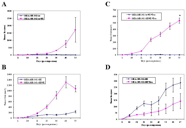
Fig. 3.

MDA-MB-361/wt cells grew tumors in nude mice with an estrogen pellet and sensitive to the growth inhibitory effects of tamoxifen, while EGFRvIII-expressing breast cancer cells were responsive to estrogen-stimulation, but were tamoxifen-resistant. Five × 106 MDA-MB-361/vIII cells were injected s.c. in ovariectomized, athymic nude mice with or without estrogen (E2) and/or tamoxifen (Tam). Four mice were used for each treatment group in this experiment, and each mouse received injections at both left and right mammary fat pads. a, MDA-MB-361/wt cells in the estrogen-treated group grew large tumors, whereas tumors that grew without an estrogen supplement were smaller and dissipating (1709.0 ± 835.3 mm3 versus 5.1 ± 3.6 mm3, p=0.2012). b, MDA-MB-361/vIII cells in the estrogen-treated group grew large tumors, whereas tumors that grew without an estrogen supplement were smaller (1068.9 ± 113.5 mm3 versus 233.0 ± 48.1 mm3, p=0.0077*). c, In the presence of estrogen and tamoxifen, MDA-MB-361/vIII cells continued to grow larger tumors than MDA-MB-361/wt cells, which had no tumor growth at the end of the treatment period (533.1 ± 39.0 mm3 versus 0 ± 0 mm3, p=0.0002*). d, EGFR/vIII cells did not have significantly reduced or enhanced tumor growth in the presence of tamoxifen alone in comparison to untreated cells (139.9 ± 59.3 mm3 versus 283.1 ± 42.3 mm3, p=0.1034). Bars, SE.
