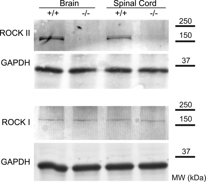
Figure 1.
Levels of ROCK protein in ROCKII−/− mice. Brain and spinal cord tissue from ROCKII−/− and wild-type mice was examined by immunoblot for ROCKII and ROCKI protein. A representative blot for each antigen is shown from one pair of three animals with similar results. Anti-GAPDH immunoblots for the same samples demonstrate equal protein loading. Migration of molecular weight (MW) standards is shown at right.

