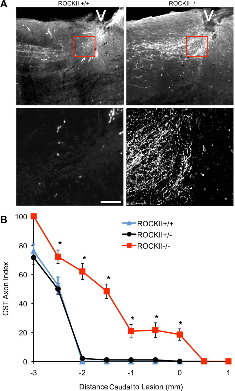
Figure 10.

ROCKII gene deletion increases the number of CST axons in the astrocytic scar of an SCI. A, Photomicrographs illustrate sagittal sections of ROCKII+/+ (left) and ROCKII−/− (right) mice 6 weeks after hemisection. BDA-immunoreactive CST axons can be seen approaching the lesion site (indicated by the white arrowheads) in both genotypes. High-power magnifications of the lesion area (highlighted boxes in top, shown in bottom) show regenerating CST axons at distances >3 mm rostral from the lesion epicenter in ROCKII+/+ mice (lower left photomicrograph), whereas ROCKII−/− CST axons are present entering the lesion site (lower right photomicrograph). Scale bar, Top, 800 μm; bottom, 200 μm. B, Quantification of CST axon growth illustrates that significantly greater numbers of axons are present at various points leading up to the lesion epicenter (mean ± SEM, 1-way ANOVA, p < 0.05) in ROCKII−/− animals (red) compared with ROCKII+/− (black) and ROCKII+/+ (gray) mice.
