
Figure 4.
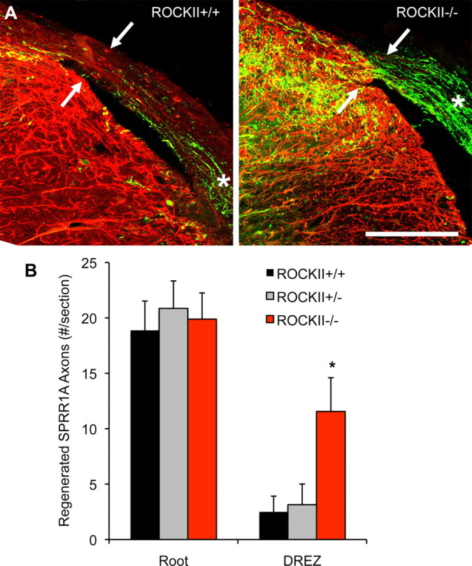
ROCKII gene deletion promotes regeneration of SPRR1A-expressing sensory neurons after rhizotomy. A, Photomicrographs illustrate high-power transverse sections of cervical spinal cord from adult ROCKII+/+ (left) and ROCKII−/− (right) mice which underwent dorsal rhizotomy at cervical spinal cord segments C5–C8 28 d previously. SPRR1A-immunoreactive axons (green channel) can be seen in the dorsal root of both genotypes (asterisk). However, significantly greater numbers of axons are present entering the DREZ in the ROCKII−/− group (arrow in right photomicrograph) than in control group (arrow in left photomicrograph, quantified in B, asterisk). Reactive CNS tissue is visualized with anti-GFAP immunofluorescence (red channel). Scale bar, 100 μm. Mean ± SEM, one-way ANOVA, p < 0.05.