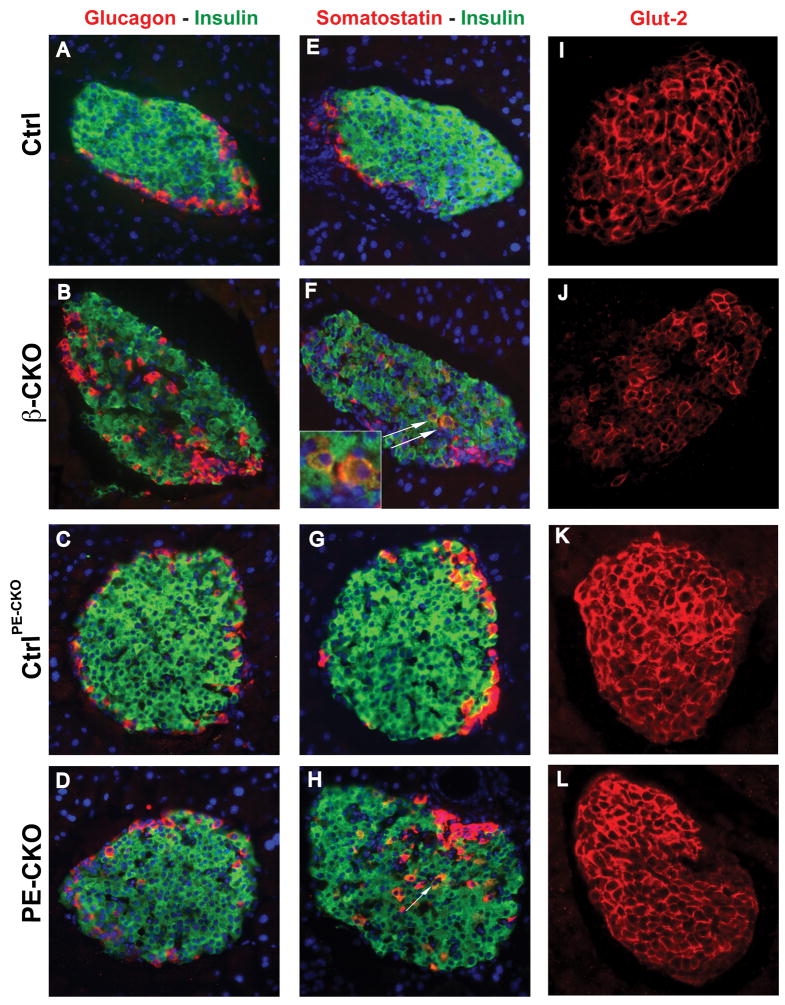
Figure 2. Effect of β cell-specific ablation of neuroD on islet characteristics.
(A–D) Pancreatic sections from control, neuroD β-CKO and neuroD PE-CKO mice were immunostained for insulin (green) and for either glucagon (red, A–D) or somatostatin (red, E–H). Nuclei are stained with DAPI (blue). The white arrows in (F) and (H) indicate insulin and somatostatin co-stained cells. Original magnification was 200x. (I–L) The expression of Glut-2 (red) in the control, neuroD β-CKO and neuroD PE-CKO pancreas. Original magnification was 200x.

