Figure 3.
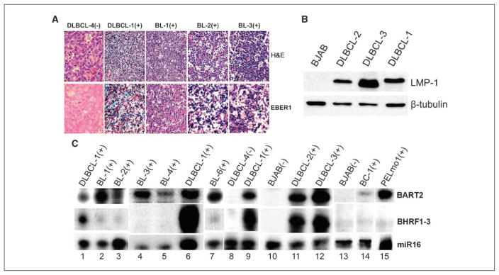
Differential expression of BHRF1-3 and BART2 miRNAs in primary unmanipulated clinical specimens. A, representative immunohistochemical analysis in primary unmanipulated clinical specimens. Endemic BLs (BL-1, BL-2, and BL-3) and primary DLBCLs (DLBCL-1 and DLBCL-4) were stained with H&E (top row) or stained for EBER1 (bottom row). Positive staining to EBV/EBER is visualized as dark blue spots. (−), EBV negative; (+), EBV positive. B, Western blot of LMP-1 protein in primary DLBCLs. EBV+ DLBCL-1, DLBCL-2, and DLBCL-3 were probed with specific antibody to LMP-1. An EBV− BL line, BJAB, was used as a negative control, and the same membrane was also probed with β-tubulin for a loading control. C, RPA analysis of BHRF1-3 and BART2 miRNA expression in primary EBV+ BLs (BL-1, BL-2, BL-3, BL-4, and BL-6), EBV+ primary DLBCLs (DLBCL-1, DLBCL-2, and DLBCL-3), a primary EBV+ PEL specimen (PELmo1), and an established EBV+ PEL line (BC-1). EBV− DLBCL-4 and BJAB were also analyzed as negative controls, and miR16 as a loading control.

