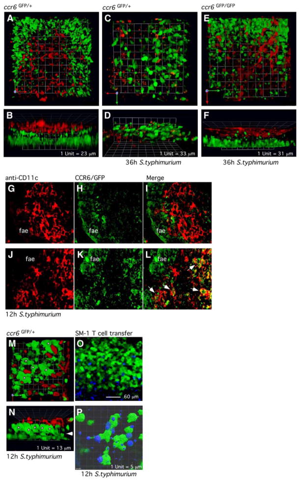
Figure 5. CCR6+ DCs Migrate into the FAE in Response to Salmonella Infection.
(A–F) Three-dimensional analysis of confocal microscopic image series from living tissues of the most distal PPs in ccr6GFP/+ and ccr6GFP/GFP mice before and after oral S. typhimurium infection. (A), (C), and (E) show 3D reconstructions viewed from the top (luminal side), and (B), (D), and (F) from the side.
(G–L) CD11c Immunofluorescence from acetone- fixed tissues of the follicle associated dome regions in ccr6GFP/+ mice before and after oral S. typhimurium infection. (G–I) Confocal images before S. typhimurium infection and (J–L) 12 hr after S. typhimurium infection. Arrows indicate recruited CCR6+CD11c+ DCs.
(M–P) Three-dimensional analysis of the dome region of PPs in ccr6GFP/+ mice after oral S. typhimurium infection. Living intestinal tissues where incubated with Rhodamine-labeled Ulex europaeus type I lectin (UEA 1) to stain M cells. Arrows indicate the basal membrane; asterisk, intraepithelial DCs. (O and P) Three-dimensional analysis of PPs in ccr6GFP/+ mice 24 hr after transfer of Cell-Tracker-blue-labeled SM-1 transgenic T cells and 12 hr after S. typhimurium infection. Quicktime movies of 3D renderings are supplied as Supplemental Data.

