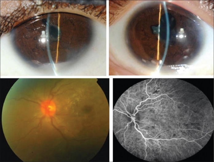
Figure 1.

Slit-lamp biomicroscopy of a 45-year-old woman with strongly positive tuberculin skin test (22 mm induration) shows mutton-fat keratic precipitates, posterior synechiae and anterior chamber fibrinous exudate (top). Fundus photograph shows optic disc swelling and hyperemia (bottom left). Indocyanine green angiography shows choroidal hypofluorescent areas (bottom left)
