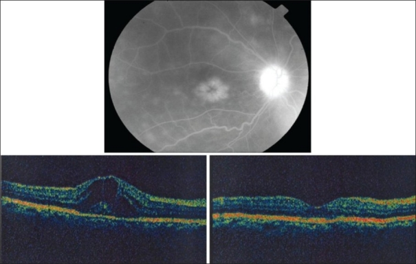
Figure 4.

Right eye of a 45-year-old woman with strongly positive tuberculin skin test (20 mm induration). Fluorescein angiography shows leakage from optic nerve head and cystoid macular edema (top). Optical coherence tomogrqaphy shows cystoid macular edema. Central macular thickness was 588 μm. Visual acuity was 20/100 (bottom left). Two months after starting antituberculous therapy and systemic corticosteroids, optical coherence tomography displays normal anatomy of the macula with reduction of central macular thickness to 239 μm. Visual acuity improved to 20/30 (bottom right)
