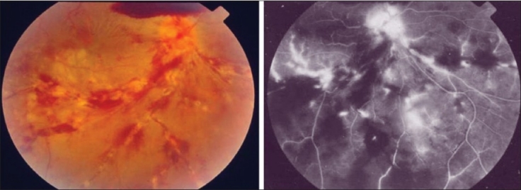
Figure 5.

The right eye of a 28-year-old man with strongly positive tuberculin skin test (20 mm induration) shows thick perivenous sheathing with intraretinal hemorrhages, cotton-wool spots, neovessels on optic nerve head, and preretinal hemorrhage above optic nerve head (left). Fluorescein angiography shows leakage from the retinal veins, and neovessels on optic nerve head and retinal nonperfusion (right)
