
Figure 6.
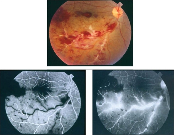
(a) Fundus photograph of the right eye of a 23-year-old man with documented Behçet's disease showing extensive perivenous sheathing involving inferotemporal vein with intraretinal hemorrhages and hemorrhagic infarction temporal to the macula (b) Fluorescein angiogram showing delayed filling of the involved vein (c) and late leakage and staining