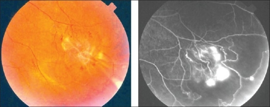
Figure 19.

Fundus photograph of a 27-year-old man with strongly positive tuberculin skin test demonstrating sclerosed white retinal vessels in the periphery and neovessels Fundus fluorescein angiogram showing peripheral capillary nonperfusion, and leakage from neovessels
