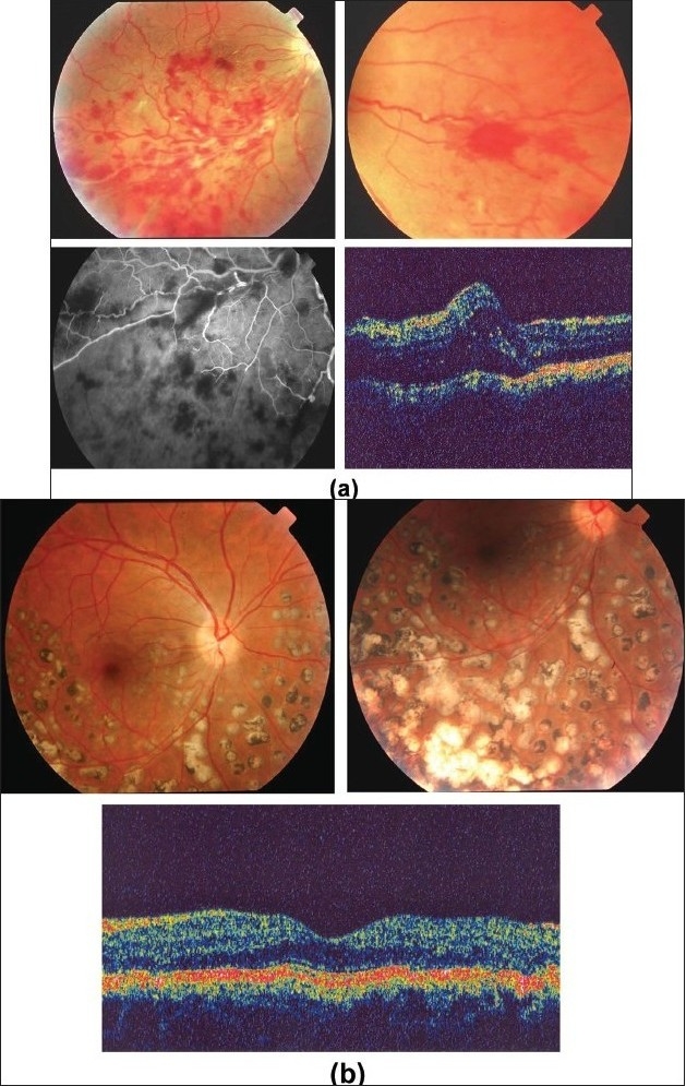
Figure 21.

(a) Fundus photographs of the right eye of a 25-year-old man with strongly positive tuberculin skin test demonstrating perivenous sheathing with intraretinal hemorrhages (upper left) and neovessels nasal to optic nerve head (upper right) Fluorescein angiography showing retinal nonperfusion (bottom left) Optical coherence tomography showing macular edema (bottom right)
(b) Fundus photographs after treatment with systemic steroids, appropriate antituberculous therapy, and scatter laser photocoagulation (upper left and right) showing resolution of perivenous sheathing and intraretinal hemorrhages and involution of neovessels Optical coherence tomography displays normal anatomy of the macula (bottom)
