
Figure 22.
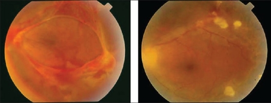
Fundus photograph of the left eye of a 32-year-old man with strongly positive tuberculin skin test showing vitreous hemorrhage and active fibrovascular tissue on the optic nerve head and along the vascular arcades (left) Fundus photograph after pars plana vitrectomy and endolaser photocoagulation (right) showing clear vitreous cavity and involution of neovessels