
Figure 23.
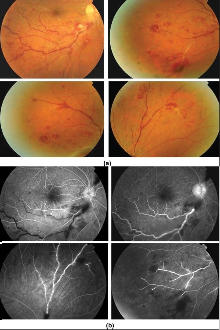
(a) Fundus photographs of the right eye of a 27-year-old man with documented Behçet’s disease showing perivenous sheathing, intraretinal hemorrhages, and retinal infiltrates
(b) Fluorescein angiogram showing delayed filling of the inferotemporal vein (upper left), late leakage and staining of the involved veins, and retinal nonperfusion (bottom left)