Figure 1.

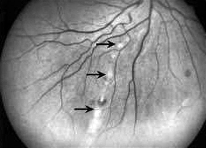
Red-free fundus photograph of the right eye of a patient with West Nile virus infection showing radial linear clustering of active deep creamy chorioretinal lesions (arrows)

Red-free fundus photograph of the right eye of a patient with West Nile virus infection showing radial linear clustering of active deep creamy chorioretinal lesions (arrows)