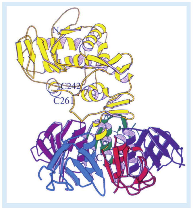
Figure 1. Ribbon diagram of Shiga toxin.

The enzymatic A-subunit (yellow) noncovalently associates with a pentameric ring of receptor-binding B-subunits. Following toxin internalization and proteolysis of the A-subunit by furin or calpain, a disulphide bond between C242 and C261 links the A1- and A2-fragments. Reproduced with permission from [3].
