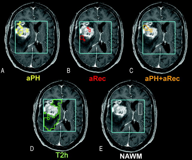
Fig 1.
Heterogeneity in the distribution of abnormal perfusion regions within a representative grade IV glioma. A, The region describing only abnormal peak height (aPH) and normal recovery patterns. B, The region consisting of only abnormal recovery (aRec) with normal peak height values. C, The intersection of both abnormal peak height and abnormal recovery (aPH+aRec) is displayed. D and E, The T2 hyperintensity lesion (T2h), which excludes all regions of abnormal perfusion, contrast enhancement, and macroscopic necrosis, and contralateral normal appearing white matter (NAWM).

