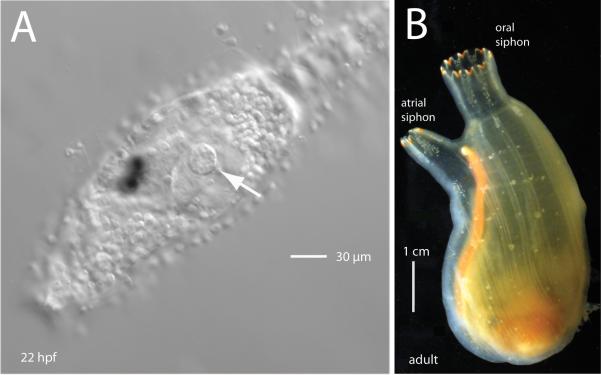
Fig.1.
The atrial primordium in Ciona development. (A) Lateral view of the head and trunk region of a Ciona savignyi larva, about 3 hours after hatching (anterior is left). The left atrial primordium is visible as a disk on the lateral epidermis (arrow). (B) An adult ascidian showing the single atrial siphon branching slightly to the left and the larger oral siphon, top center. The oral siphon arises from a single rudiment, whereas the adult atrial siphon results from the fusion of paired juvenile atrial siphons.

