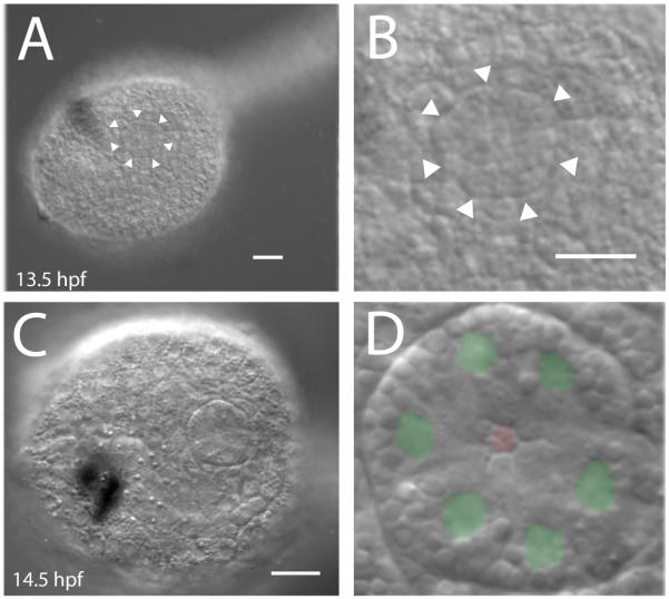
Fig. 2.
(A) First appearance of the atrial primordium in Ciona tailbud embryos. At about 13 hours post fertilization the Ciona tail begins to straighten. About 30 minutes later, a distinct circular cluster of cells with a clearly defined border appears in the epidermis. (B) Detail of lateral trunk epidermis in A. (C) DIC image showing surface epidermis of recently formed atrial primordium. (D) Detail of C with green where nuclei are observed and a red hexagon at the central aperture of the atrial primordium. Scale bar = 20 μm.

