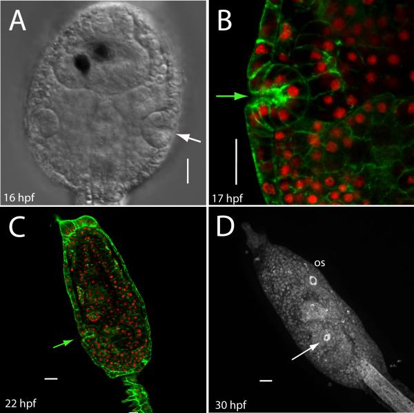
Fig. 3.
Atrial invagination occurs soon after primordium formation. (A) At about 16 hpf, a dorsal view shows an invaginated primordium with incipient atrium. Anterior is up. Pigmented cells within the sensory vesicle are visible, central midline canal is dorsal nervous system; notochord is not visible in this view. (B) An optical section through a larval primordium reveals a shallow atrium (F-actin is green and nuclei are red). (C) Confocal section of a Ciona larva after head elongation. Arrow shows atrial aperture. (D) Fluorescent label reveals that the mature larva has a ring of actin at the atriopore. Arrow points to the atrial aperture; a similar arrangement of actin is found at the oral siphon opening (os). Scale bar = 20 μm.

