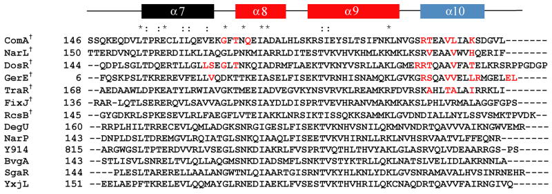
Figure 1.
Sequence alignment of the C-terminus of competence protein A (ComAC) with other members of the NarL family of proteins. The secondary structure prediction is shown on top. Red coloring of the secondary structure represents the helix-turn-helix domain and blue represents the helix involved in the dimerization interface. Residues in red highlight those residues identified as being specifically involved in the dimerization interface. *denotes conserved residues, : denotes conserved substitutions, †denotes proteins with known structures. The known structures include: ComAC (current work), NarL (PDB 1JE8) 35, DosR (PDB 1ZLJ) 36, GerE (PDB 1FSE)37, RcsB (PDB 1P4W)38, FixJC (1X3U)39, and TraR (1H0M and 1L3L)40.

