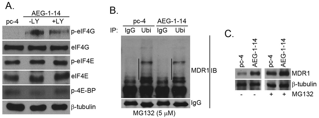
Fig. 5.
AEG-1 augments translation initiation and decreases degradation of MDR-1. A. The expression of the indicated proteins were detected in Hep-pc-4 (pc-4) cells and Hep-AEG-1-14 (AEG-1-14) cells treated or untreated with 10 µM LY294002 for 24 h by Western blot analysis. B. Hep-pc-4 (pc-4) and Hep-AEG-1-14 (AEG-1-14) cells were treated with MG132 (5 µM) for 24 h and the cell lysates were subjected to immunoprecipitation by anti-ubiquitin antibody (Ubi). The immunoprecipitates were subjected to Western blot analysis using anti-MDR1 antibody. IgG: normal mouse IgG. The lines in the figure indicate ubiquitinated MDR1. C. Hep-pc-4 (pc-4) and Hep-AEG-1-14 (AEG-1-14) cells were treated with MG132 (5 µM) for 24 h and the expression of MDR1 and β-tubulin was analyzed by Western blot.

