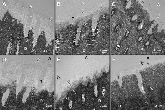
Figure 3.

Transmission electron micrographs of unstained, non-demineralized sections of Adper Prompt L-Pop-bonded dentin slabs after biomimetic remineralization. Generic abbreviations: A, adhesive; T, dentinal tubule; D, mineralized intertubular dentin. (A) A 15-second-etched control specimen depicting the resin-dentin interface before remineralization. The aggressive self-etch adhesive created a 4- to 5-µm-thick zone of completely demineralized dentin (between open arrows). (B) A 15-second-etched specimen after 3 mos of remineralization. A 3- to 4-µm-thick remineralized zone (between open arrowheads) could be identified along the base of the original hybrid layer (between open arrows). The top of the original hybrid layer was probably better infiltrated by adhesive resin and did not remineralize because it was free of water-filled voids. (C) A 15-second-etched specimen after 6 mos of remineralization. Only the base (between open arrowheads) of the original hybrid layer (between open arrows) was remineralized. (D) A 60-second-etched control specimen revealing a 9- to 10-µm-thick zone of demineralized dentin. (E) A 60-second-etched specimen after 3 mos of remineralization. Partial remineralization had occurred along the basal 5 µm of the original 10-µm-thick hybrid layer (between open arrows). (F) A 60-second-etched specimen after 6 mos of remineralization. The remineralized base of the hybrid layer was 9 µm thick (between open arrowheads) and was almost as dense as the underlying naturally mineralized dentin. Despite such heavy remineralization in the basal portion, the superficial 2 µm of the original hybrid layer (between open arrows) did not remineralize. Collectively, the basal locations of the remineralized hybrid layers corresponded to the locations where the hybrid layers disappeared after prolonged aging of the specimen slabs in glycine buffer.
