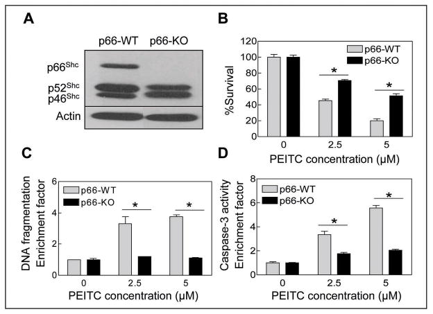
Fig. 1.
A, immunoblotting for p66Shc protein using lysates from untreated MEFs derived from wild-type (p66-WT) and p66Shc knockout mice (p66-KO). The blot was stripped and re-probed with anti-actin antibody to ensure equal protein loading. Cell viability (B), cytoplasmic histone-associated DNA fragmentation (C), and caspase-3 activation (D) in p66-WT and p66-KO MEFs following 4 h treatment with DMSO (control) or the indicated concentrations of PEITC. Results are expressed as enrichment factor relative to DMSO-treated control. Each experiment was done at least twice in triplicate and representative data from a single experiment are shown. Columns, mean (n=3); bars, SE. *Significantly different (P<0.05) between the indicated groups by paired t-test.

