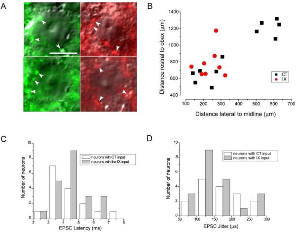
Fig. 1.
Characterization of monosynaptic contacts between gustatory afferent input and second order rNST neurons. A. Examples of DIC images of rNST neurons surrounded by labeled afferent profiles. The profiles surrounding the neurons on the left resulted from Alexa Fluor dextran 488 (green) applied to the CT and the profiles surrounding the neurons on the right resulted from Alexa Fluor dextran 568 (red) applied to the IX nerve. Examples of the labeled profiles are indicated by white arrow heads. Bar = 10 μm. B. Location of a subset of the recorded neurons. Lucifer yellow labeled neurons with definite co-ordinates were mapped relative their position rostral to the obex and lateral to the midline of the brainstem. C Distribution of synaptic latencies evoked by ST stimulation for neurons with input from the CT and IX nerves. D. Distribution of the standard deviation of synaptic latency (jitter) for neurons with input from the CT and IX nerves.

