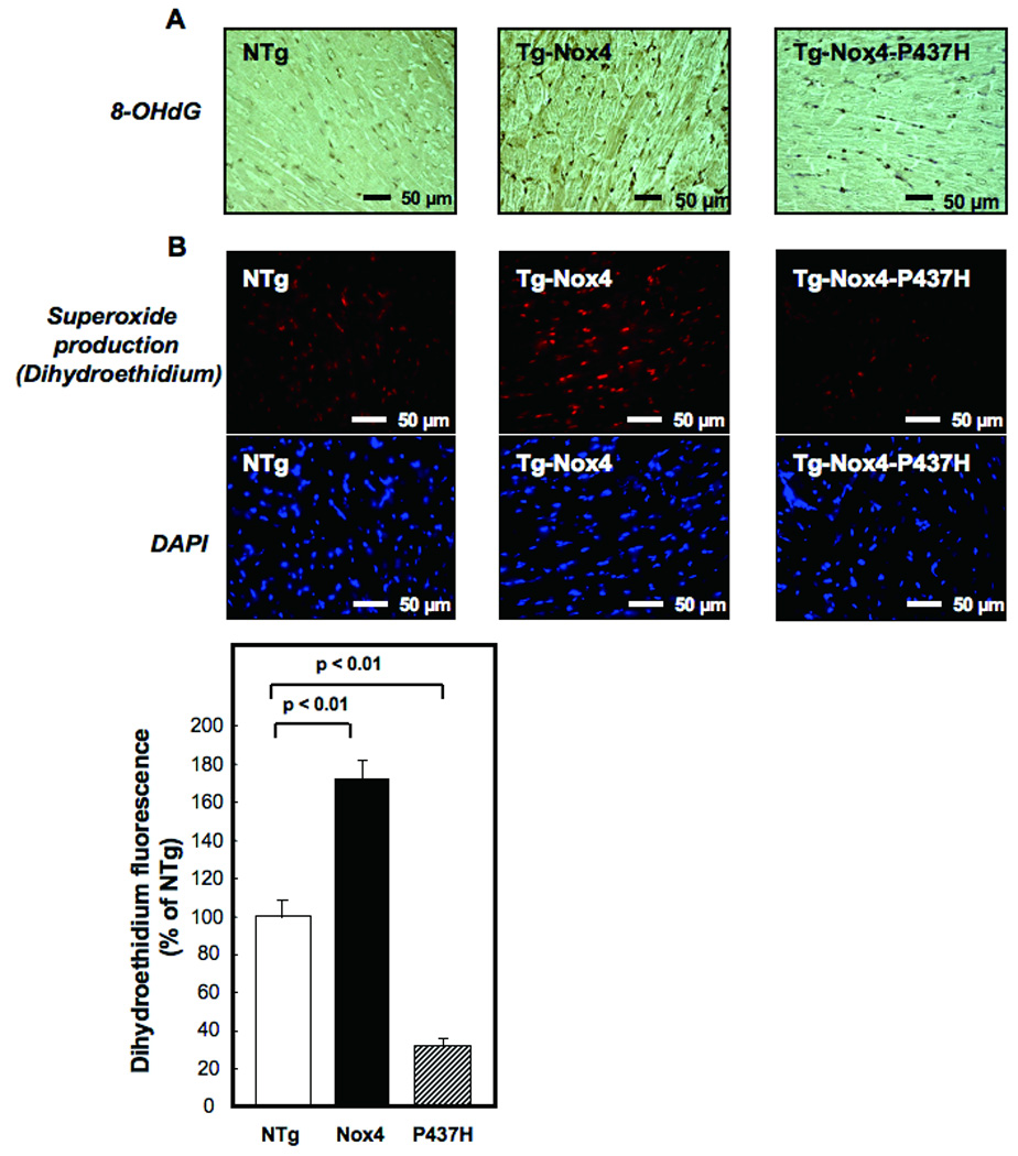
Figure 3.

The extent of oxidative stress in the LV was determined by 8-hydroxyl-deoxyguanosine (8-OHdG) staining (A) and dihydroethidium (DHE) staining (B). In B, DAPI staining shows nuclei. LV sections were prepared from Tg-Nox4, Tg-Nox4-P437H and NTg mice.
