
Figure 5.
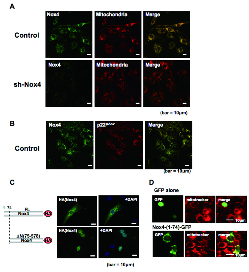
Subcellular localization of Nox4 in cardiac myocytes. A) Cultured cardiac myocytes were transduced with adenovirus harboring shRNA-scramble or shRNA-Nox4. Myocytes were co-stained with anti-Nox4 antibody and anti-F0F1 ATP synthase antibody, a marker of mitochondria. Merged images are shown on the right. B) Cultured cardiac myocytes were co-stained with anti-Nox4 and anti-p22phox antibodies. A merged image is shown on the right. C) Cultured cardiac myocytes were transduced with an expression plasmid harboring full length (FL) Nox4-HA or Nox4 lacking MLS (ΔN(75–578))-HA. Forty-eight hours after transfection, cells were stained with anti-HA antibody and DAPI. Truncation of the N-terminal region (1–74) causes disappearance of the perinuclear staining of Nox4 in cardiac myocytes. D) Cardiac myocytes were transduced with expression plasmids harboring either GFP alone or Nox4 (1–74)-GFP. Representative images of GFP, Mitotracker and merged images are shown.