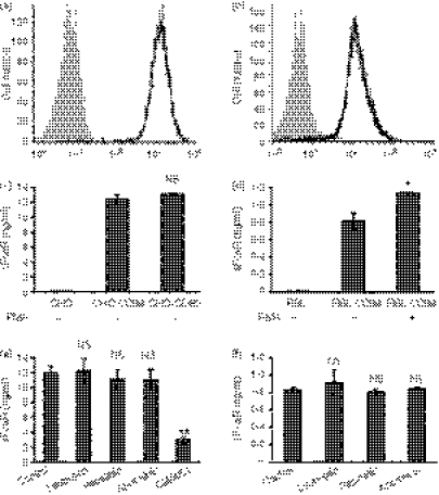
Figure 3.
FcαR shedding from stable transfectants of Chinese Hamster ovary (CHO) cells and rat basophilic leukaemia (RBL) cells. (a) and (b) Flow cytometry analysis of FcαR stably expressed on CHO (a) and RBL (b) cells. CHO or RBL cells (filled histogram) and CHO-CD89 or RBL-CD89 cells (black line) were incubated with fluorescein isothiocyanate (FITC)-conjugated MIP8a-F(ab′)2 for 60 min on ice, then washed and analysed using flow cytometry. (c) and (d) FcαR shedding from CHO and RBL cells stably expressing FcαR. A total of 2 × 10⁵ cells were cultured with or without phorbol 12-myristate 13-acetate (PMA) (10 ng/ml) for 24 hr, and the concentration of soluble FcαR (sFcαR) in the supernatants was determined using enzyme-linked immunosorbent assay (ELISA). (e) and (f) CHO-CD89 cells or RBL-CD89 cells were cultured in the presence of leupeptin (2 μg/ml), pepstatin (2 μg/ml), aprotinin (2 μg/ml) or GM6001 (20 μm) for 24 hr. The concentration of sFcαR in the supernatants was determined using enzyme-linked immunosorbent assay (ELISA) analyses. Data are the mean ± standard error of the mean (SEM) of three independent experiments. *P <0·01, **P <0·001. NS, not significant.

