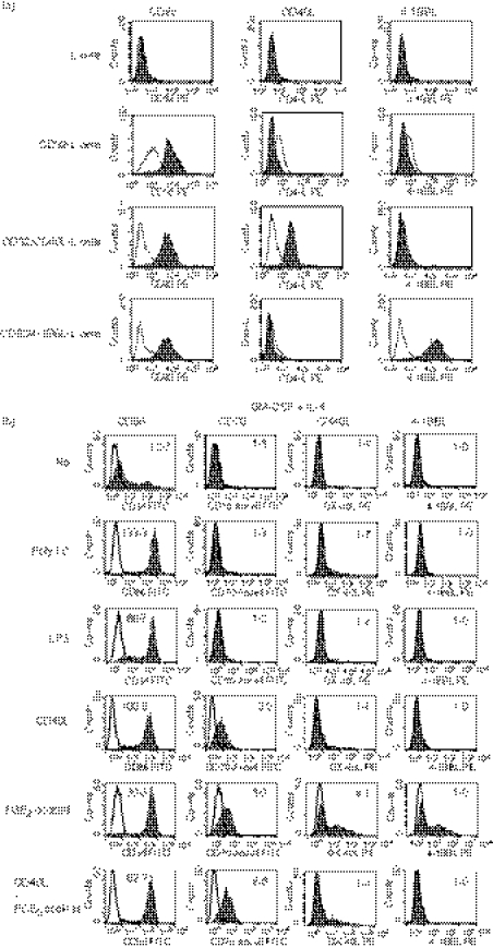
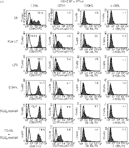
Figure 1.
Expression of CD70, OX40 ligand (OX40L), 4-1BBL and CD86 on monocyte-derived dendritic cells (MoDCs) cultured in different conditions. (a) Parental L cells and those transfected with CD32 (CD32-L), CD32 and CD40L (CD32/CD40L-L) or CD32 and 4-1BBL (CD32/4-1BBL-L) were stained with phycoerythrin (PE) -labelled anti-CD32, anti-CD40L or anti-4-1BBL monoclonal antibody (mAb). Open histograms represent cells stained with isotype-matched control mAbs. (b, c) Monocytes were cultured for 3 days in R10 in the presence of interleukin-4 (IL-4) (b) or interferon-α (IFN-α) (c) together with granulocyte–macrophage colony-stimulating factor (GM-CSF) to induce immature IL-4-DCs or IFN-DCs, respectively. Thereafter, they were cultured for 3 days in the absence or presence of maturation-inducing stimuli [poly I:C, lipopolysaccharide (LPS), CD40L-L cells, or the prostaglandin E2 (PGE2) cocktail]. The cells were stained with fluorescein isothiocyanate (FITC) -labelled anti-CD86, anti-CD70 (clone BU69), PE-labelled anti-OX40L, or anti-4-1BBL mAb. Open histograms represent cells stained with isotype-matched control mAbs. The numbers shown with each histogram represent [mean fluorescence intensity (MFI) of each costimulatory molecule]/(MFI of isotype-matched control]. Data are representative of three experiments.


