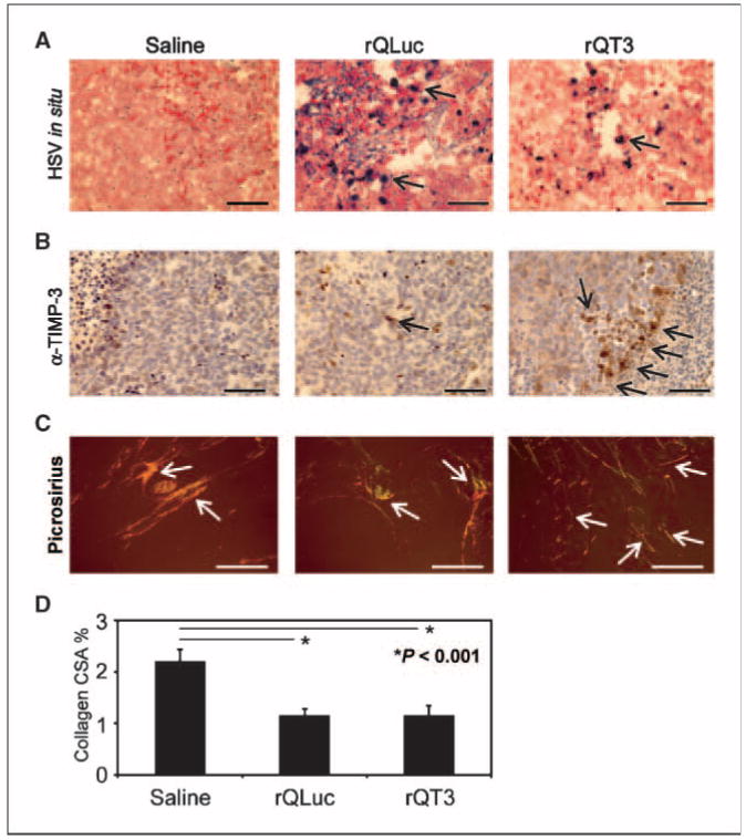
Figure 4.

Neuroblastoma xenografts injected with oHSV showed virus infection, TIMP-3 expression, and a disrupted microenvironment. A, ∼500 mm3 neuroblastoma (LA-N-5) xenografts, at 72 h posttreatment with multiple doses of saline, rQLuc, or rQT3 at day 21 (see Fig. 6A), were examined for HSV-1 infection by in situ hybridization. Only virus-treated tumors show positive HSV-1 staining. B, TIMP-3 expression in tumors treated with rQT3 showed positive cells in clusters compared with only occasional, scattered cells in saline- or rQLuc-treated tumors. Magnification, ×400; scale bars, 50 μm. Regions of necrosis/apoptosis are evident by the karyorectic cells (dark nuclei) in the periphery of the images of saline- and rQT3-treated tumors. C, saline-treated tumors stained with picrosirius red showed thick, highly structured collagen illustrated by orange and yellow birefringence under polarized light. Magnification, ×100; scale bars, 250 μm. oHSV-treated tumors showed less structured collagen indicated by green and blue birefringence. D, quantification of tumor collagen cross-sectional area (CSA) or % birefringent area.
