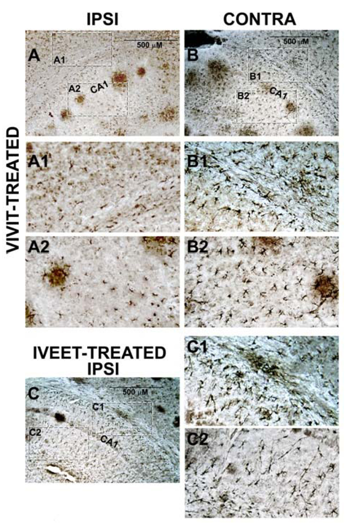
Figure 2. The NFAT inhibitor, VIVIT, reduces astrocyte activation in AD mice.
A and B, photomicrographs of a GFAP-labeled coronal section from a 14-month-old APP/PS1 mouse treated intraventricularly for two weeks with 11R-VIVIT. Dashed boxes indicate regions of higher magnification shown in A1 and A2 (ipsilateral, VIVIT-treated hemisphere—Ipsi) and B1 and B2 (contralateral hemisphere—contra). C, GFAP-labeled coronal section (Ipsilateral hemisphere) from a 14-month-old APP/PS1 mouse treated for two weeks with 11R-IVEET control peptide. Dashed boxes indicate regions of higher magnification shown in C1 and C2. IVEET-treated mice and the contralateral hemisphere of VIVIT-treated mice appear to express more numerous and more ramified astrocytes relative to the ipsilateral hemisphere of VIVIT-treated mice. CA1 = cornu ammonis 1 pyramidal cell layer.

