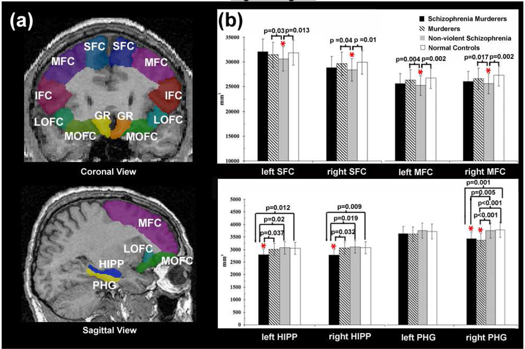
Figure 1.
(a) Illustrations of the segmentation of the frontal cortex of one subject on the coronal (top) and sagittal view (bottom). SFC: superior frontal cortex, MFC: middle frontal cortex, IFC: inferior frontal cortex, lOFC: lateral orbitofrontal cortex, mOFC: medial orbitofrontal cortex, GR: gyrus rectus, HIPP: hippocampus, PHG: parahippocampal gyrus. (b) Gray matter volumes of the left and right SFC and MFC (top) and HIPP and PHG (bottom) in schizophrenia murderers, murderers, non-violent schizophrenia patients, and normal controls. P values indicate significant group comparisons while controlling for whole brain volume. The vertical lines represent the standard error bars.

