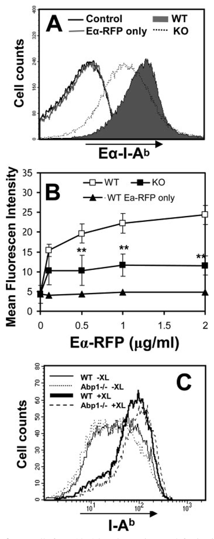
FIGURE 3.
B cells from Abp1 knockout mice are defective in Ag processing and presentation. A, Splenic B cells from wt (filled) and Abp1−/− (dotted) mice were incubated at 37°C for 10 min with EαRFP (1 µg/ml) alone (gray line) or with the Ab complex that targets EαRFP to the BCR. Cells were washed and incubated at 37°C for 14 h. Eα peptide-loaded MHC class II I-Ab complexes on the cell surface were detected using anti-Y-Ae mAb and quantified using flow cytometry. B, Splenic B cells from wt (□) and Abp1−/− (■) mice were incubated with different concentrations of EαRFP alone or EαRFP-Ab complexes, as described in A. Splenic B cells from wt mice were incubated with different concentrations of EαRFP alone (▲). Cells were stained and analyzed, as described above. Shown are the averages (±SD) of mean fluorescence intensity of Y-Ae staining from three independent experiments. **, p < 0.01. C, The expression levels of MHC class II I-Ab of splenic B cells from wt (solid lines) and Abp1−/− (dotted lines) mice before (−XL) and after (+XL) exposure to EαRFP-Ab complexes were measured using flow cytometry.

